Technology is the set of techniques, instruments and systems used to discover, describe and explain different phenomena. The pulmonary hypertension research group has developed and optimized several technologies to understand the disease and to discover treatments aimed at improving the quality of life of patients.
We divide these different techniques into two categories: basic research and clinical research.

Inverted microscopy
The laboratory is equipped with a high-performance inverted microscope (Axio Observer, Zeiss) with a module for 3D sectioning and an incubation chamber. It allows the analysis of several types of stains.
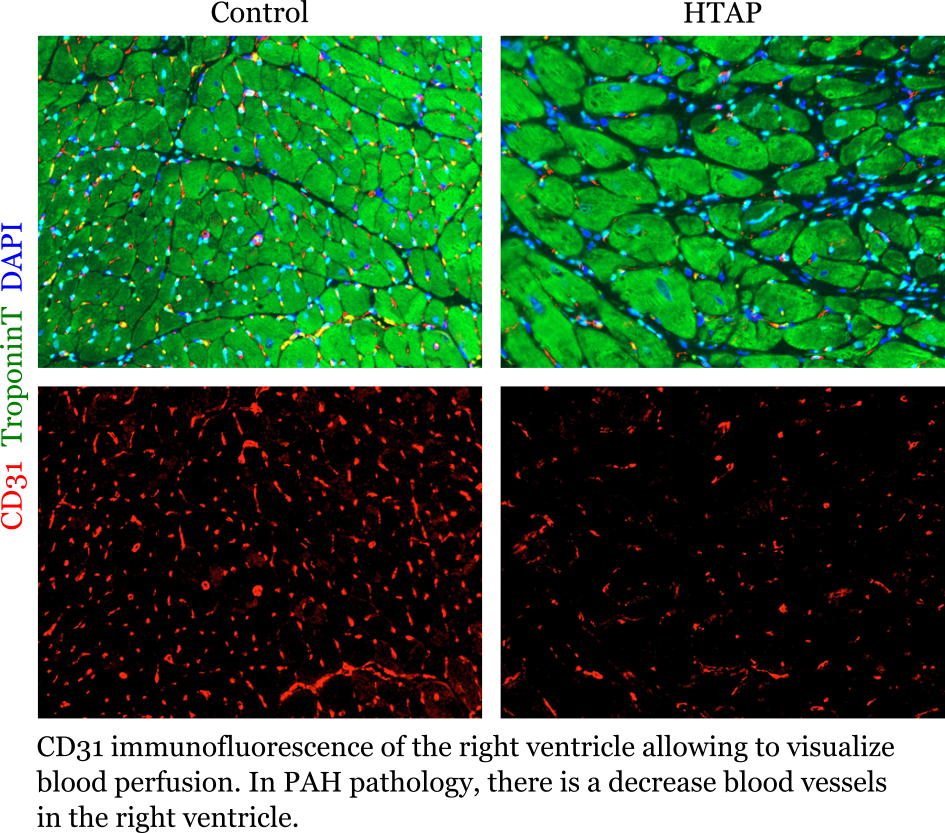
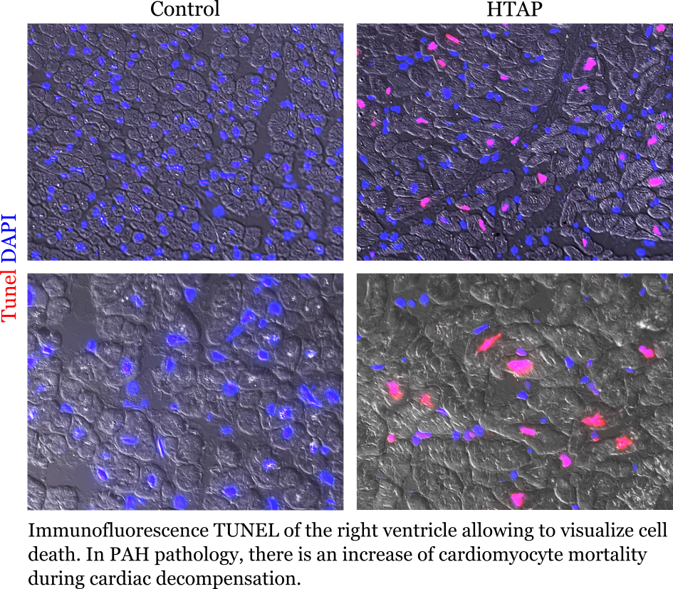
Immunofluorescence histology
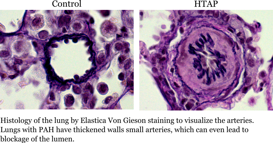
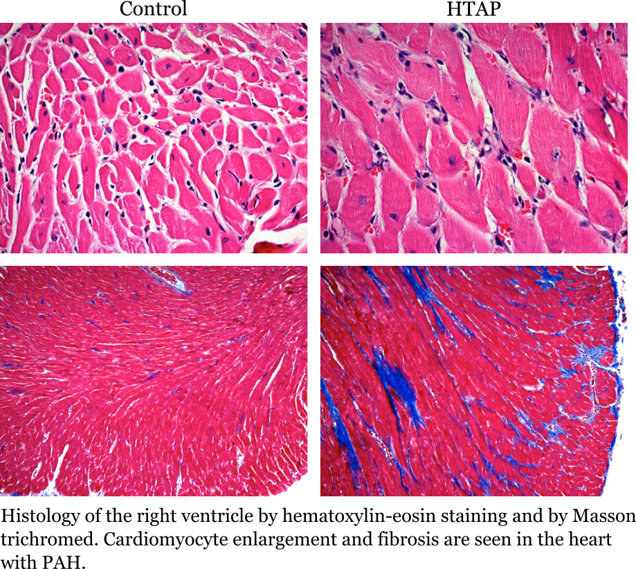
Histology by colorimetry
ChemiDoc
The ChemiDoc is an imaging system for the detection of chemiluminescence. It is used for the visualization of proteins isolated from biopsies or cells in culture.

qRT-PCR
Real-time PCR is a technique that involves amplifying DNA fragments in order to quantify them. This method, widely used in research, requires DNA from a few cells in culture or from a biopsy and provides important information for the identification of potential therapeutic targets.
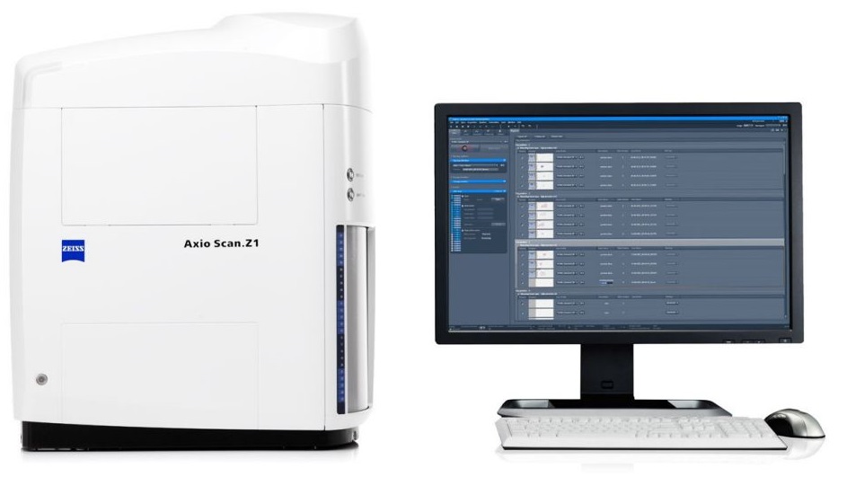
Zeiss AxioScan.Z1 slide scanner
The Zeiss AxioScan.Z1 slide scanner enables automated scanning of up to 100 microscope slides (brightfield or fluorescence) into high quality virtual slides.
Cellular culture
The use of human cells isolated from patients with pulmonary hypertension is essential for research. The identification of potential therapeutic targets requires molecular and epigenetic analyzes of these cells.

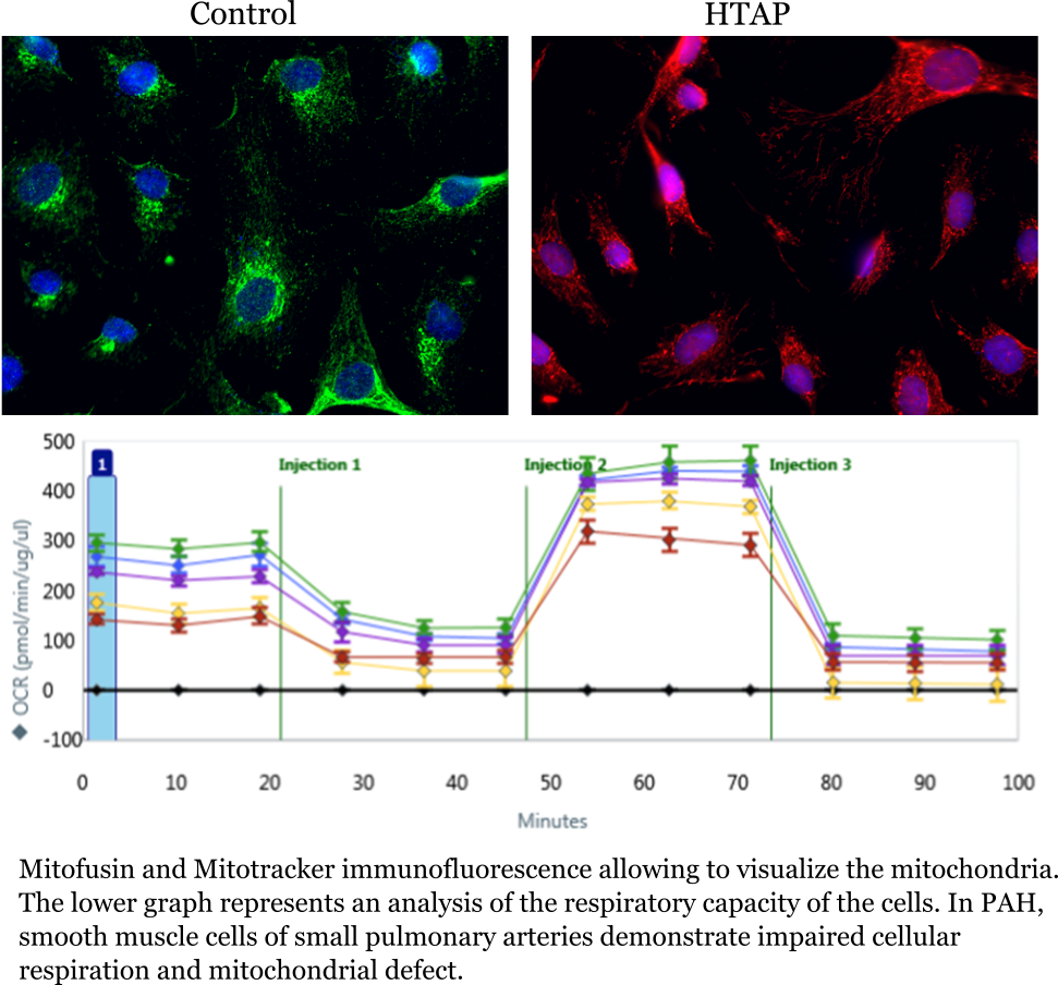
Seahorse XFe24 technology
This technology allows the measurement of oxygen consumption and extracellular acidification, two important indicators for cellular respiration and glycolysis. Combined, this information tells us about the metabolic functions of cells.
Histology unit
The histology unit is a platform belonging to the pulmonary hypertension research group and available to the entire research center. It includes all the devices necessary for specimen fixation, paraffin embedding and cutting for microscopic analysis. For more information on the histology unit, to know the prices or to make a reservation, contact us at groupe.recherche.htap@criucpq.ulaval.ca
Available devices
- Excelsior AS
- PrintMate AS
- HistoStar AS
- SlideMate AS
- Microtome

Tomography
Tomography is an imaging technique using X-rays that allows us to learn a lot about the physiology of the disease.

Cardiac catheterization
Cardiac catheterization is an essential test for the diagnosis of pulmonary hypertension. It consists of inserting a pressure and volume sensor into the pulmonary artery. This procedure is used both clinically by the doctor for the diagnosis of the disease, and in basic research to verify the effectiveness of the treatments under study.
BiStim magnetic stimulator
Magnetic stimulation of the femoral nerve causes an involuntary contraction of the quadriceps. The force of voluntary contraction of the quadriceps is compared to the force of involuntary contraction generated by this device, which is used as a maximum reference measurement. The BiStim is directly connected to an amplifier and an analysis module.
Biodex System 4 pro
This robotic and computerized dynamometer makes it possible to measure muscle strength by applying different types of loads (isometric, isokinetic, isotonic). In research, it is mainly used to assess the strength and endurance of the quadriceps, but its applications are wide (post-injury rehabilitation, identification of the risk of falling, neurorehabilitation, etc.)
Non-ferromagnetic ergocycle
description to come
Ergocycle with electromagnetic brake
Combined with a respiratory analysis module (Vmax 29 spectra), the ergocycle is mainly used in the clinical laboratory to determine cardiopulmonary capacity. However, it can be used for a multitude of exercise protocols such as a constant load endurance test or VO2 Max test.
Portable telemetry system for respiratory physiology assessment
The portable nature of this device makes it possible to measure the gas exchanges of each breath while the subject is moving. It can therefore monitor several physiological parameters (heart rate, VO2, VCO2, minute ventilation, saturation, etc.) continuously during outpatient tests.
Near-infrared spectroscopy (NIRS)
This system measures the variation in muscle and / or cerebral oxygenation and the variation in blood volume. We can also get muscle oxygen saturation data and muscle oxygen consumption.
Non-invasive continuous heart monitoring
Digital arterial pulse plethysmography allows constant measurement of arterial pressure and advanced hemodynamic variables.
Electromyography
Placing the electromyograph electrode on a muscle measures electrical activity there. This makes it possible to determine the muscles recruited, their degree of activation and the order in which they are recruited.
Complete analysis of respiratory functions
The Vmax Encore with plethysmography module allows you to perform tests such as spirometry, diffusion capacity, measurement of lung volumes, lung compliance as well as inspiratory and expiratory pressure. It is also possible to measure total lung capacity by removing nitrogen with the Platinum Body Plethysmograph.
Muscle biopsy
This surgical procedure is performed under local anesthesia by an experienced doctor. A thin incision allows the insertion of a biopsy trocar which collects a muscle sample. This is then analyzed in the fundamental laboratory.